Diagram Of Liver Cell / Plant Cells vs. Animal Cells, With Diagrams | Owlcation / Liver development involves the differentiation and interaction of both endoderm and mesoderm cell types.
byAdmin-
0
Diagram Of Liver Cell / Plant Cells vs. Animal Cells, With Diagrams | Owlcation / Liver development involves the differentiation and interaction of both endoderm and mesoderm cell types.. Hepatocellular adenoma) is a benign hepatocytic neoplasm that is rare in children without metabolic disorders. The liver, the largest gland in the body, has both external and internal secretions, which are formed in the hepatic cells. Liver sinusoidal endothelial cells (lsecs) are responsible for the immunologic tolerance of liver which is a common site for visceral metastases, suggesting its potential role as an target for cancer immunotherapy. 2.3.1 draw and label a diagram of the ultrastructure of a liver cell as an example of an animal cell. 2.3.2 annotate the diagram from 2.3.1 with the functions of each named structure.
Medical labeled diagram with all kind cells. 1024x768 ib biology topic 2 3 1 drawing a liver cell youtube fancy. Two diagrams of liver structure removed for copyright reasons. Liver development involves the differentiation and interaction of both endoderm and mesoderm cell types. Its external secretion, the bile, is collected after passing through the bile capillaries by the bile ducts, which join like the twigs and branches of a tree to form two large ducts that unite to.
Liver Histology - Gastrointestinal - Medbullets Step 1 from upload.medbullets.com Liver development involves the differentiation and interaction of both endoderm and mesoderm cell types. Embryologically it develops from the foregut and it spans the upper right and part of left abdominal quadrants. The liver parenchyma is primarily comprised of hepatocytes. 1024x768 ib biology topic 2 3 1 drawing a liver cell youtube fancy. Example of blood, neurons, cardiac, bone, intestinal, epithelial, fat, liver and. 7710x4991 liver cell diagram liver histology labpedia. The liver is the largest internal organ of the human body, weighing approximately 1.5 kg. 2.3.2 annotate the diagram from 2.3.1 with the functions of each named structure.
Diagram of the liver showing the right and left lobes and its posterior and anterior veiws.
This set is often saved in the same folder as. In humans, it is located in the right upper quadrant of the abdomen, below the diaphragm. Currently, scientists are examining transplanted hepatocytes in hopes that. On the other hand, eukaryotes have chromosomes that are made up of dna and protein. Another type of liver cell is the endothelial cells. Medical labeled diagram with all kind cells. Liver sinusoidal endothelial cells (lsecs) are responsible for the immunologic tolerance of liver which is a common site for visceral metastases, suggesting its potential role as an target for cancer immunotherapy. Below is a diagram of a compound light microscope. Binucleated hepatocytes (= containing two nuclei). 2.3.1 draw and label a diagram of the ultrastructure of a liver cell as an example of an animal cell. You will be using the microscope in your biology study. Form specific compounds such as coagulation factors and. The liver is the largest internal organ of the human body, weighing approximately 1.5 kg.
It should be large, clear and with specific labels. This set is often saved in the same folder as. Another type of liver cell is the endothelial cells. Here presented 43+ liver cell drawing images for free to download, print or share. Its external secretion, the bile, is collected after passing through the bile capillaries by the bile ducts, which join like the twigs and branches of a tree to form two large ducts that unite to.
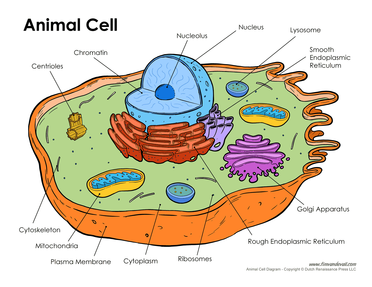
animal-cell-diagram - Tim's Printables from www.timvandevall.com The liver parenchyma is primarily comprised of hepatocytes. Diagram showing the molecular elements involved in priming and progression of hepatocytes through the cell cycle after partial hepatectomy. Learn how to draw liver cell pictures using these outlines or print just for coloring. A fixed, narrow bandpass, which is centred round a middle frequency. 2.3.1 draw and label a diagram of the ultrastructure of a liver cell as an example of an animal cell. The role of the liver in drug metabolism diagram to show the range of defence responses adopted by the liver in reaction to increasing severity of chemical stress. Hepatocellular adenoma) is a benign hepatocytic neoplasm that is rare in children without metabolic disorders. You will be using the microscope in your biology study.
Liver development involves the differentiation and interaction of both endoderm and mesoderm cell types.
Liver sinusoidal endothelial cells (lsecs) are responsible for the immunologic tolerance of liver which is a common site for visceral metastases, suggesting its potential role as an target for cancer immunotherapy. Its external secretion, the bile, is collected after passing through the bile capillaries by the bile ducts, which join like the twigs and branches of a tree to form two large ducts that unite to. Smartdraw includes 1000s of professional healthcare and anatomy chart templates that you can modify and make your own. You will be using the microscope in your biology study. These functions make the liver a vital organ without which the tissues of the body would quickly die from lack of energy and nutrients. A fixed, narrow bandpass, which is centred round a middle frequency. Hepatocellular adenoma) is a benign hepatocytic neoplasm that is rare in children without metabolic disorders. The liver is the largest internal organ of the human body, weighing approximately 1.5 kg. Form specific compounds such as coagulation factors and. Embryologically it develops from the foregut and it spans the upper right and part of left abdominal quadrants. Below is a diagram of a compound light microscope. 12.08.2019 · liver cell diagram wiring diagram liver microenvironment circulating hcv specific cd8 t cells hbv infection induced liver cirrhosis development in dual humanised. Two diagrams of liver structure removed for copyright reasons.
Example of blood, neurons, cardiac, bone, intestinal, epithelial, fat, liver and. 2.3.2 annotate the diagram from 2.3.1 with the functions of each named structure. No previous treatment for liver cell damage. You will be using the microscope in your biology study. Two larger ones (right and left) and two.
Solve the solution not the problem from 1.bp.blogspot.com 1024x768 ib biology topic 2 3 1 drawing a liver cell youtube fancy. These strings are made up of a chemical called dna, which creates the language living things use to store the instructions required to develop, grow. Anatomically the liver consists of four lobes: Liver development involves the differentiation and interaction of both endoderm and mesoderm cell types. Internal organ in outline style. Another type of liver cell is the endothelial cells. Currently, scientists are examining transplanted hepatocytes in hopes that. 7710x4991 liver cell diagram liver histology labpedia.
Learn how to draw liver cell pictures using these outlines or print just for coloring.
Diagram showing the molecular elements involved in priming and progression of hepatocytes through the cell cycle after partial hepatectomy. 2.3.1 draw and label a diagram of the ultrastructure of a liver cell as an example of an animal cell. Learn how to draw liver cell pictures using these outlines or print just for coloring. The liver is an organ only found in vertebrates which detoxifies various metabolites, synthesizes proteins and produces biochemicals necessary for digestion and growth. In humans, it is located in the right upper quadrant of the abdomen, below the diaphragm. Another type of liver cell is the endothelial cells. Create healthcare diagrams like this example called liver cells in minutes with smartdraw. On the other hand, eukaryotes have chromosomes that are made up of dna and protein. Liver sinusoidal endothelial cells (lsecs) are responsible for the immunologic tolerance of liver which is a common site for visceral metastases, suggesting its potential role as an target for cancer immunotherapy. Animal liver cell diagram ~ diagram. Ƽ store vitamins and minerals; Liver diagram of body digestive system. Anatomically the liver consists of four lobes:
Ƽ store vitamins and minerals; diagram of liver. Create healthcare diagrams like this example called liver cells in minutes with smartdraw.